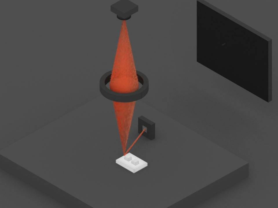
Transmission Electron Microscope Working Animation
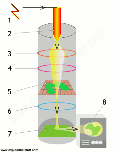
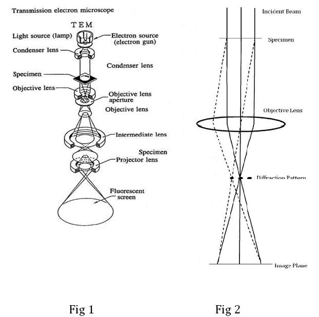
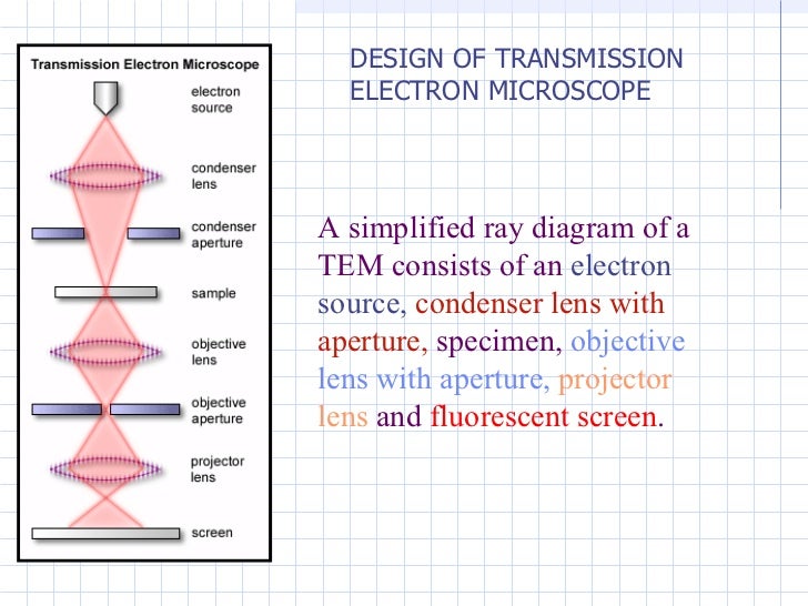
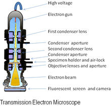
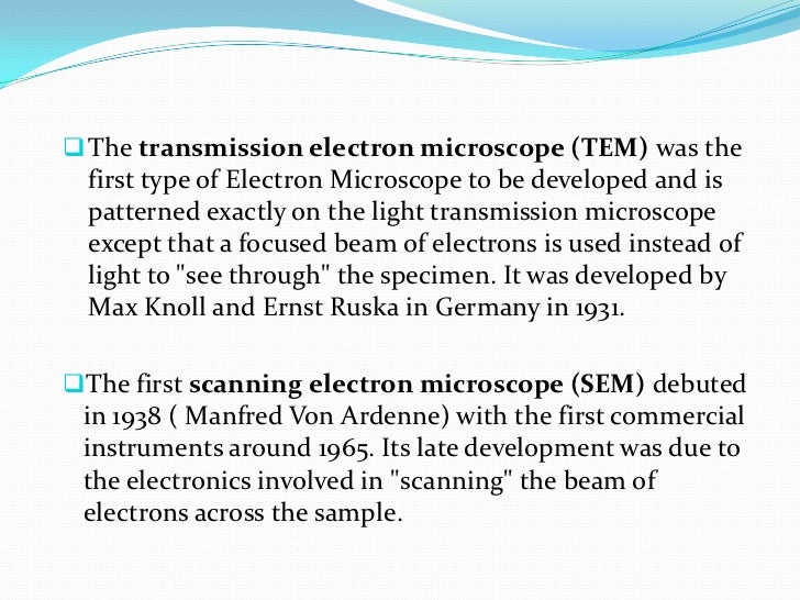
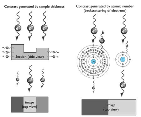
Transmission electron microscopy tem is a microscopy technique in which a beam of electrons is transmitted through a specimen to form an image.
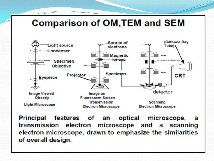
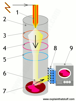
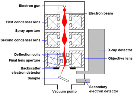
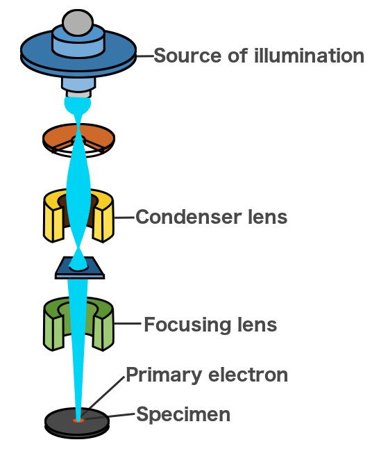
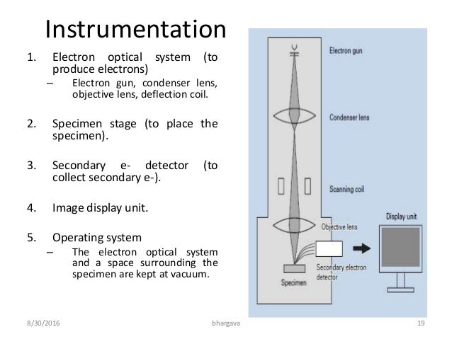
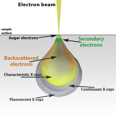
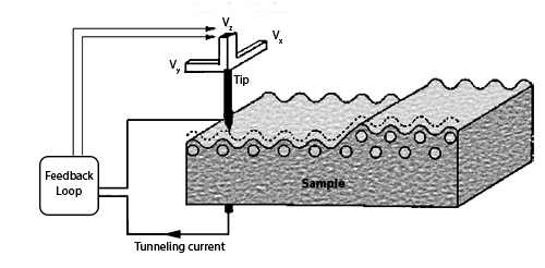
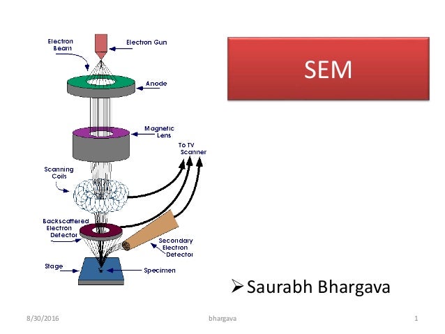
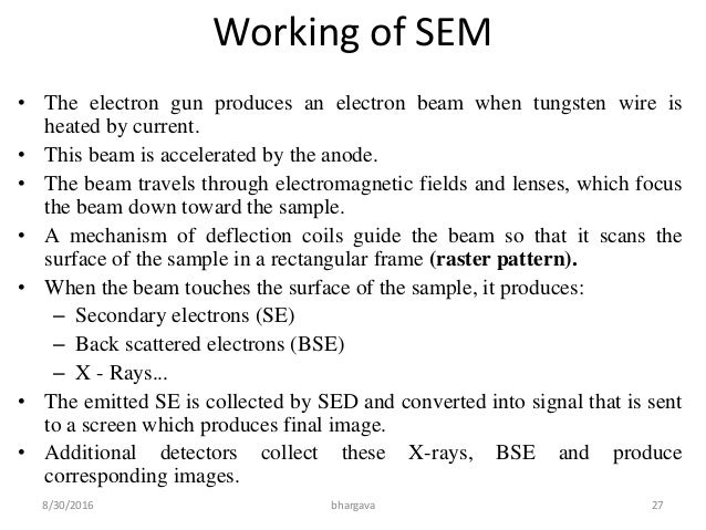
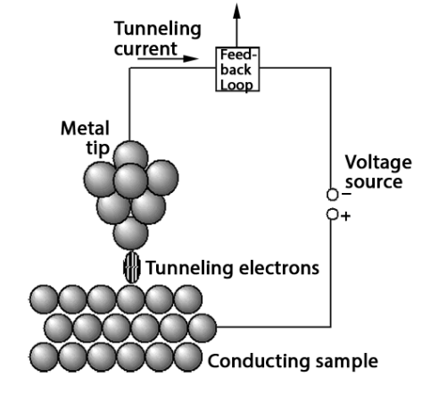
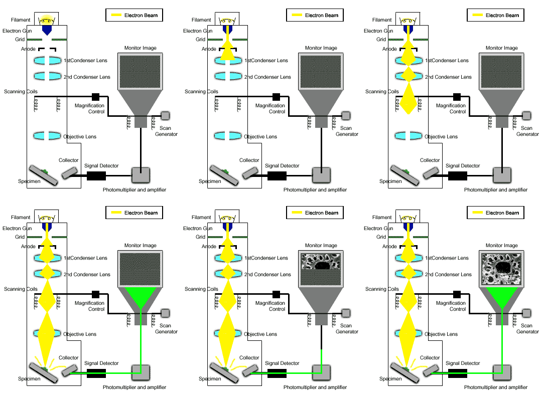
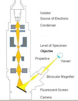
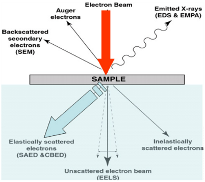
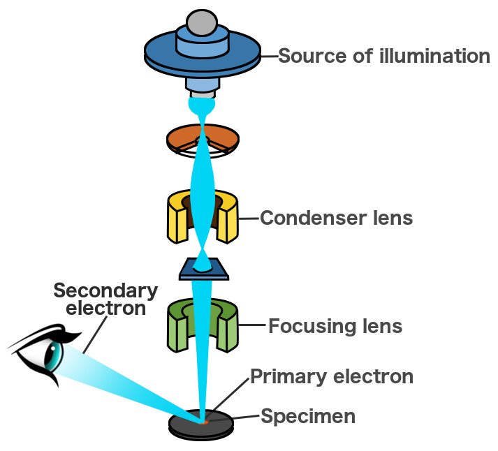
Transmission electron microscope working animation. There are 3 types of electron microscopes namely the transmission electron microscope tem scanning electron microscope sem and scanning tunneling microscope stm. Read this article to learn about the working principle of electron microscopes with diagram. An image is formed from the interaction of the electrons with the sample as the beam is transmitted through the specimen. After understanding that an accelerated electron beam can have a very high resolving power we then move on to see how one can use this electron beam in imaging technology.
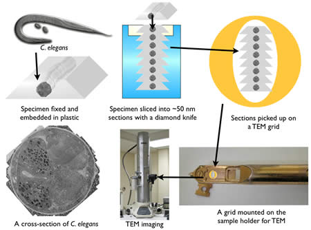
Imaging in the light microscope and the transmission electron microscope. The specimen is most often an ultrathin section less than 100 nm thick or a suspension on a grid. A microscopy technique where an electron beam transmits through an ultra thin specimen and a nanometer scale shadow image is taken. Transmission electron microscopy tem is used to assess formation of the fibrils predominantly by measuring fibril diameter.
3 function phctf or short ctf is discussed. Here we describe an enhanced protocol for measuring fibril diameter as well as fibril volume fraction mean fibril length fibril cross sectional shape and fibril 3d organization that are also major determinants of. Atomic world transmission electron microscopetem animations download animation download animation lotus effect transmission electron microscope scanning tunneling microscope carbon nanostructures eulers formula social issues eulers formula social issues. In particular emphasis is placed on the basic concepts of phase contrast microscopy.
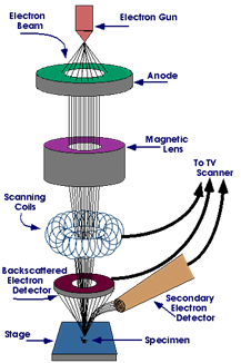
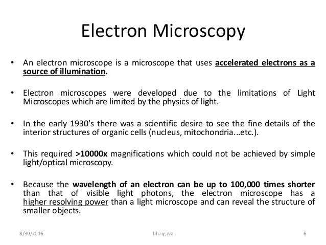
An electron microscope uses an electron beam to produce the image of the object and magnification is obtained by electromagnetic fields. To familiarize the technique of sample preparation for transmission electron microscopy. Electron beams are used in electron microscope to illuminate the specimen and thus creates an image. To familiarize the technique of sample preparation for transmission electron microscopy.
